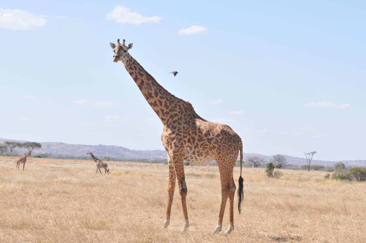
- Giraffe skin disease, a mystery condition that inflicts crusty lesions on the world’s tallest animal, has been recorded in 13 giraffe populations in seven African countries. It is particularly widespread in Tanzania.
- Researchers used camera trap images to quantify how severe the disease was among giraffe populations in Tanzania’s Serengeti and Ruaha national parks.
- They found that most cases of the infections that the camera traps detected were “mild” or “moderate” according to a scale they devised, suggesting that the disease, although widespread, is likely not life-threatening at the moment.
- The researchers have, however, observed that giraffes with more severe infections tend to move with difficulty, which could make them more vulnerable to lion predation — a hypothesis they are now investigating with data from Ruaha National Park.
Giraffes have many problems to deal with. There’s habitat loss and poaching. Then there’s a mysterious skin disease that’s been recorded in 13 giraffe populations in seven African countries.
The condition, termed simply the giraffe skin disease, starts off as small nodules on the animal’s skin. The nodules can develop into dry, scaly patches, which then turn into large crusty, grayish-brown lesions filled with blood or pus. Researchers are only beginning to wrap their heads around the little-understood disease. Arthur Muneza, a doctoral student at Michigan State University and East Africa coordinator of the Giraffe Conservation Foundation, is one of them.

Muneza has been studying the disease in Tanzania, where it’s widespread, affecting the giraffe’s long limbs. About a quarter of the giraffes in Serengeti National Park show signs of the disease. In Tarangire National Park, some 63 percent of giraffes suffer from it, while in Ruaha National Park, around 86 percent of the giraffe population sport the characteristic skin lesions.
“To go to an area and see almost all the animals with signs of this disease is quite surprising,” Muneza told Mongabay.
The high prevalence of the disease in Tanzania sparked a question in Muneza’s mind. How severe is the infection among Tanzania’s giraffes?
A few studies have tried to answer this question in the past, but their classifications of giraffe skin disease severity were very subjective, Muneza said. “There is no standard way of defining what a mild giraffe skin disease is, what moderate giraffe skin disease is, and what constitutes severe giraffe skin disease.”
To develop a classification that’s less arbitrary, Muneza and his colleagues turned to camera traps in a new study published in the Journal of Wildlife Diseases.
The researchers selected all giraffe photos that had been captured during extensive camera trap surveys in Ruaha and Serengeti, then whittled down the list to those that showed the full extent of all four legs of the giraffes, from shoulder joints to hooves.
The team then used photogrammetry techniques on the final 405 photos to quantify giraffe skin disease in the animals. They measured the vertical length of both the lesions and the legs visible in the photos, determined the proportion of the leg that was affected by the lesions, then statistically grouped these numbers to get three different categories of lesion severity.
Giraffes with less than 16 percent of their legs covered by lesions were classified as having mild giraffe skin disease, those with 16 to 25 percent of the legs covered had a moderate form of the disease, and individuals with more than a quarter of their leg covered by lesions had severe skin disease, according to the study.
“At the moment, we know very little about the disease, so our assumption is that the external and physical manifestation of the disease is the indicator we can use to categorise the severity of the disease,” Muneza told Mongabay.
While the cameras usually captured photos of giraffe limbs, it’s the animals’ upper bodies that have unique coat patterns. This meant that the researchers couldn’t identify individual animals from the camera trap photos alone. This could potentially bring in bias if the disease severity classifications had been estimated from multiple photos of just a few giraffes. To see if this was the case, the researchers compared the severity rates estimated by the camera trap images with those obtained from another technique: photographs of 305 individually recognized giraffes that the researchers had taken using digital cameras during vehicle-based surveys in both parks. Both techniques produced similar results.
The photos revealed that lesions of giraffe skin disease were, in general, more common on the front legs of the animals than the back legs. Moreover, most cases of the disease that the camera traps in Ruaha and Serengeti detected were considered “mild,” followed by “moderate” forms.
“What this means is that there’s no need to overreact at the moment, and that the disease is not as severe as we would like to think,” Muneza said. “Externally it looks uncomfortable, it looks bad. But it’s still the mild and moderate forms of the disease.”
Previously, researchers relied on close observations of the animals to describe the severity of giraffe skin disease. But such a technique is not only laborious, it also limits the spatial extent one can cover, Muneza and his colleagues write in the paper. Camera traps, on the other hand, are “noninvasive, can be rapidly deployable, and are applicable to a variety of species,” they add.
Miranda Sadar, an assistant professor at Colorado State University, who’s been investigating the cause of giraffe skin disease and was not involved in the study, said that photogrammetry is indeed becoming a more widely used tool “to monitor the size of injuries in animals in a non-invasive way.”
Not everyone is convinced, however.
“This was a laudable attempt to use camera trap images to quantify giraffe skin disease, but the method is significantly less useful than active observations,” Monica Bond, a wildlife biologist at the U.S.-based Wild Nature Institute who studies giraffes in Tanzania and was not involved in the study, told Mongabay.
Observing giraffes directly is easy, she said, because the animals are calm and vehicles can drive up close to them, allowing easy examination using binoculars. This way, researchers can also identify giraffes individually, and inspect the animals’ bodies from multiple angles to understand how and where the disease has spread, Bond added. The camera trap images at the moment don’t allow for both individual identification and a thorough whole-body examination of the giraffes, she said.
Muneza agreed that camera trap images have some challenges. For example, the data set would be more useful had the cameras been placed higher. “We could have used the camera trap data to identify individual giraffes, and that could have given us a more robust dataset to quantify the categories of giraffe skin disease,” he said. “We were able to use only use photos of the legs.”
The lesions also don’t always appear on the legs. In Uganda, for example, giraffes more commonly get infections on their neck and shoulders.

Muneza’s study, however, adds to the growing evidence that giraffe skin disease is probably not life-threatening for the animals in the studied parks — at least for now. Bond, too, in a study published in 2016, found that giraffes with lesions in Tanzania’s Tarangire National Park had similar survival rates as those without lesions.
That doesn’t mean that researchers should stop studying the disease, Muneza said.
His team has observed, for instance, that giraffes with more severe infections tend to move with difficulty, which could make them more vulnerable to lion predation. The researchers are now investigating this hypothesis with data from Ruaha National Park.
Muneza’s team is also working with Tanzanian authorities to figure out what exactly causes giraffe skin disease. Some preliminary studies suggest that a filarial worm could be transmitting the disease, with secondary fungal infections worsening it. But researchers are yet to pin down the actual causative agent and how the disease spreads.
“One of the biggest concerns is that if it is a filarial worm, then we need to see if it crosses over to cattle,” Muneza said. “Given that you have communities that live near parks, and some of them graze their cattle near and around the giraffe areas, there is potential for the disease to cross over to livestock. And if it does, that will then affect the perceptions that people have towards sharing landscapes with wildlife, which is a big challenge in East Africa.”
Citation:
Muneza, A. B., Ortiz-Calo, W., Packer, C., Cusack, J. J., Jones, T., Palmer, M. S., … Montgomery, R. A. (2019). Quantifying the severity of giraffe skin disease via photogrammetry analysis of camera trap data. Journal of Wildlife Diseases, 55(4), 770-781. doi:10.7589/2018-06-149